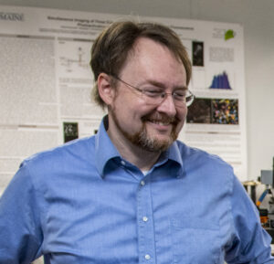
Samuel T. Hess
 Samuel T. Hess
Professor of Physics
Graduate Coordinator
- B.S., Physics, Yale University, 1995
- M.S., Physics, Cornell University, 1998
- Ph.D., Physics, Cornell University, 2002
Office: 313 Bennett Hall
Phone: 207.581.1036
Email: samuel.hess@maine.edu
um.grad.physics@maine.edu
Other Webpages:
- Research Page
- IMB Webpage
- FPALM Instrumentation Development Collaborations
- Hess Lab Advisees and Alumni 6
Research Interests:
- Experimental and Theoretical Biophysics,
- Fluorescence Microscopy and Spectroscopy,
- Function and Lateral Organization of Biomembranes,
- Single Molecule Fluorescence Photophysics,
- Green Fluorescent Proteins.
Recent Presentations:
“Super-Resolution Microscopy: Illuminating Biological Functions to Cure Disease,” Science Seminar Series, Emera Astronomy Center, University of Maine, Orono, ME, September 2019.
“Localization Microscopy: Technological Advances Coupled to Biological Applications” Keynote Lecture, Molecular Biophysics Training Program, Cornell University, Ithaca, NY, November 2019.
“Super-Resolution Microscopy Quantifies Interactions between Influenza Hemagglutinin, Actin, and PIP2,” Department of Chemistry, Yale University, New Haven, CT, May 2019.
“Localization Microscopy: Methodological Advances for Biological Applications,” Gordon Research Conference “Single Molecule Approaches to Biology,” Mt. Snow, Vermont, July 2018.
“The Role of Probe Photophysics in Localization-Based Superresolution Microscopy,” Biophysical Society Annual Meeting, San Francisco, CA, February 2018.
“Localization Microscopy: Technological Advances Coupled to Biological Advances,” National Institute of Biomedical Imaging and Biotechnology, National Institutes of Health, Bethesda, MD, November 2016.
“Localization-Based Superresolution Microscopy: Principles and Biological Applications,” Edinburgh Super-Resolution Imaging Consortium (ESRIC) Summer School, Edinburgh, Scotland, August, 2016.
“Localization-Based Superresolution Microscopy: Biological Applications,” lecture for the International School of Biophysics Antonio Borsellino, 43rd Course: Nanoscale biophysics: focus on methods and techniques, Directed by A. Diaspro and P. Bianchini, Erice, Sicily, April 21, 2016.
“Localization-Based Superresolution Microscopy: Principles,” lecture for the International School of Biophysics Antonio Borsellino, 43rd Course: Nanoscale biophysics: focus on methods and techniques, Directed by A. Diaspro and P. Bianchini, Erice, Sicily, April 19, 2016.
Recent publications:
Slattery, Lucinda K., Zackery B. McClelland, and Samuel T. Hess. 2024. “Process–Structure–Property Relationship Development in Large-Format Additive Manufacturing: Fiber Alignment and Ultimate Tensile Strength” Materials 17, no. 7: 1526.
https://doi.org/10.3390/ma17071526
Sandberg, Amanda L., Avery C. S. Bond, Lucas J. Bennett, Sophie E. Craig, David P. Winski, Lara C. Kirkby, Abby R. Kraemer, Kristina G. Kelly, Samuel T. Hess, and Melissa S. Maginnis. 2024. “GPCR Inhibitors Have Antiviral Properties against JC Polyomavirus Infection” Viruses 16, no. 10: 1559. https://doi.org/10.3390/v16101559
“Conserved sequence features in intracellular domains of viral spike proteins,” Vinh-Nhan Ngo, David P. Winski, Brandon Aho, Pauline L. Kamath, Benjamin L. King, Hang Waters, Joshua Zimmerberg, Alexander Sodt, Samuel T. Hess, Virology, Volume 599, 2024, 110198, ISSN 0042-6822,
https://www.sciencedirect.com/science/article/pii/S0042682224002198
“Phosphatidylinositol 4,5-Bisphosphate Mediates the Co-Distribution of Influenza A Hemagglutinin and Matrix Protein M1 at the Plasma Membrane,” P. Raut, B. Obeng, H. Waters, J. Zimmerberg, J. Gosse, and S.T. Hess, Viruses 14(11):2509 (2022).
“Dynamics and Patterning of 5-Hydroxytryptamine 2 Subtype Receptors in JC Polyomavirus Entry,” K. Mehmood, M.P. Wilczek, J.K. DuShane, M.T. Parent, C.L. Mayberry, J.N. Wallace, F.L. Levasseur, T.M. Fong, S.T. Hess, and M.S. Maginnis, Viruses 14(12): 2597 (2022).
“Localization-Based Super-Resolution Microscopy Reveals Relationship between SARS-CoV2 Spike and Phosphatidylinositol (4,5)-bisphosphate,” P. Raut, H. Waters, J. Zimmberberg, B. Obeng, J. Gosse, and S.T. Hess, Proc. of SPIE Vol. 11965: 1196503, 1605-7422 (2022).
“Cetylpyridinium chloride (CPC) reduces zebrafish mortality from influenza infection: Super-resolution microscopy reveals CPC interference with multiple protein interactions with
phosphatidylinositol 4,5-bisphosphate in immune function,” P. Raut, S.R. Weller, B. Obeng, B.L. Soos, B.E. West, C.M. Potts, S. Sangroula, M. S. Kinney, J.E. Burnell, B.L. King, J.A. Gosse, and S.T. Hess, Toxicology and Applied Pharmacology, 440: 115913 (2022).
“Triclosan disrupts immune cell function by depressing Ca 2+ influx following acidification of the cytoplasm,”Sangroula, S., Vasquez, A.Y.,Raut, P., Obeng, B., Shim, J.K., Bagley, G.D., West, B.E., Burnell, J.E., Kinney, M.S., Potts, C.M., Weller, S.R., Kelley, J.B., Hess, S.T., Gosse, J.A.* Toxicology and Applied Pharmacology 405: 115205, doi: 10.1016/j.taap.2020.115205, 2020. PMID: 32835763
“Detection and Analysis of Uncharged Particles Utilizing Consumer-Grade CCDs,” John A. Cummings, James W. Deaton, Charles T. Hess, Samuel T. Hess,Health Physics Journal118(6):583-592 (2020).
“Quantification of Mitochondrial Membrane Curvature by Three-Dimensional Localization Microscopy,” Matthew Parent and Samuel T. Hess,iScience Notes,4(3): 1-2 (2019).
“Influenza Hemagglutinin Modulates Phosphatidylinositol(4,5)bisphosphate (PIP2) Clustering,” Nikki M. Curthoys, Michael J. Mlodzianoski, Matthew Parent, Prakash Raut, Michael B. Butler, Jaqulin Wallace, Jennifer Lilieholm, Kashif Mehmood, Melissa Maginnis, Hang Waters, Brad Busse, Joshua Zimmerberg, and Samuel T. Hess,Biophysical Journal116(5):893-909. doi: 10.1016/j.bpj.2019.01.017 (2019).
“Molecular Imaging with Neural Training of Identification Algorithm (MINuTIA),” A.J. Nelson and S.T. Hess,Microscopy Research and Technique(2018).
“Antimicrobial Agent Triclosan Disrupts Mitochondrial Structure, Revealed by Super-resolution Microscopy, and Inhibits Mast Cell Signaling via Calcium Modulation Toxicology and Applied Pharmacology,” Lisa M Weatherly, Andrew J. Nelson, Juyoung Shim, Abigail M. Riitano, Erik D. Gerson, Andrew J. Hart, Jaime de Juan-Sanz, Timothy A. Ryan, Roger Sher, Samuel T. Hess, Julie A. Gosse,Toxicololgy and Applied Pharmacology349: 39-54 (2018).
“Total internal reflection fluorescence based multiplane localization microscopy enables super-resolved volume imaging,” P.P. Mondal, S.T. Hess,Applied Physics Letters110 (21), 211102 (2017).
“The Role of Probe Photophysics in Localization-Based Superresolution Microscopy,” Francesca Pennacchietti, Travis Gould, and Samuel T. Hess,Biophysical Journal113 (9) 2037–2054 (2017).
“A Cross Beam Excitation Geometry for Localization Microscopy,” Matthew Valles and Samuel T. Hess,iScience NotesDOI: http://doi.org/10.22580/2016/iSciNoteJ2.2.1 (2017).
“Spectral Fluorescence Photoactivation Localization Microscopy,” Michael Mlodzianoski, M. S. Gunewardene, and Samuel T. Hess,PLoS One11(3): e0147506 (2016).
“Clean Localization Super-resolution Microscopy for 3D Biological Imaging,” Partha P. Mondal, Nikki M. Curthoys, and Samuel T. Hess,AIP Advances6, 015017 (2016).
“Super Resolution Fluorescence Localization Microscopy,” Michael J. Mlodzianoski, Matthew M. Valles, Samuel T. Hess, inEncyclopedia of Cell Biology, Ed. Ralph Bradshaw, Philip Stahl and John Heuser & Sergio Grinstein (2016).
“Spectral Fluorescence Photoactivation Localization Microscopy,” Michael Mlodzianoski, M. S. Gunewardene, and Samuel T. Hess, PLoS One 11(3): e0147506 (2016).
“Clean Localization Super-resolution Microscopy for 3D Biological Imaging,” Partha P. Mondal, Nikki M. Curthoys, and Samuel T. Hess, AIP Advances 6, 015017 (2016).
“Super Resolution Fluorescence Localization Microscopy,” Michael J. Mlodzianoski, Matthew M. Valles, Samuel T. Hess, in Encyclopedia of Cell Biology, Ed. Ralph Bradshaw, Philip Stahl and John Heuser & Sergio Grinstein (2016).
“Antimicrobial Agent Triclosan is a Proton Ionophore Uncoupler of Mitochondria in Living Rat and Human Mast Cells and in Primary Human Keratinocytes,” Lisa M. Weatherly, Juyoung Shim, Hina N. Hashmi, Rachel H. Kennedy, Samuel T. Hess, and Julie A. Gosse, Journal of Applied Toxicology Jul 23. doi: 10.1002/jat.3209 (2015).
“Dances with Membranes: Breakthroughs from Super-Resolution Imaging,” Nikki M. Curthoys, Matthew Parent, Michael Mlodzianoski, Andrew J. Nelson, Jennifer Lilieholm, Michael B. Butler, Matthew Valles, and Samuel T. Hess, in Current Topics in Membranes, Ed. Anne Kenworthy (2015).
“Nanoscale Imaging of Caveolin-1 Membrane Domains in vivo,” Kristin A. Gabor, Dahan Kim, Carol H. Kim, and Samuel T. Hess, PLoS One 10(2): e0117225 (2015).
“Combining Total Internal Reflection Sum Frequency Spectroscopy Spectral Imaging and Confocal Fluorescence Microscopy,” Edward S. Allgeyer, Sarah M. Sterling, Mudalige S. Gunewardene, Samuel T. Hess, David J. Neivandt, and Michael D. Mason, Langmuir 31 (3): 987-994 (2015).
“Precisely and accurately localizing single emitters in fluorescence microscopy: state-of-the-art and best practice,” Hendrik Deschout, Francesca Cella Zanacchi, Michael Mlodzianoski*, Alberto Diaspro, Joerg Bewersdorf, Samuel T. Hess, and Kevin Braeckmans, Nature Methods, accepted.
